 Magnetic resonans imaging (MRI) is an imaging technique used for high quality images of the body's organs and soft parts. Magnetic resonans imaging (MRI) is an imaging technique used for high quality images of the body's organs and soft parts.
The human body is primarily fat and water. Fat and water have many hydrogen atoms which make the human body approximately 63% hydrogen atoms. Protons in the hydrogen atoms possess a property called spin, which can be thought of as a small magnetic field.
Magnetic resonance imaging is based on the absorption and emission of energy in the radio frequency range of the electromagnetic spectrum and uses the spatial variations in the energy's phase and frequency from the illustrated object. By means of the spectroscopic MR technique it is possible to get chemical microscopic information about the molecules.
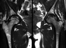
Traditional x-ray images show bone tissue, not cartilage tissue. It is often the quality and the quantity of cartilage tissue that decide whether joint preservation or joint compensatory surgery is possible. Therefore, the MR technique gives important information to orthopaedic surgeons.
When performing joint preservation operations such as Ganz osteotomy for the treatment of hip dysplasia, it is advantageous to examine the thickness of the cartilage in the articulated surfaces. This should be done before the operation and in the years following the operation in order to estimate whether the development of osteoarthritis continues.
MRI does not cause radiation impact, which is a great advantage, but because of the magnetic resonance not all patients can go through this examination and orthopaedic surgical osteosyntheses or prosthesis can make up a contra indication for MR. |